The Scientist
Born at the lab bench.
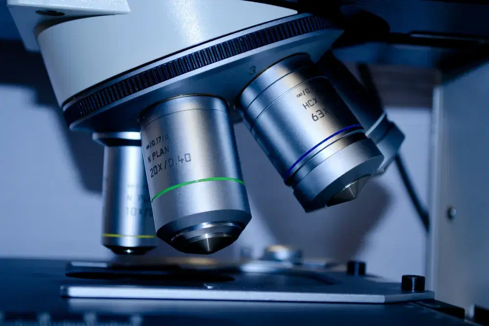
Micrographer is the work of a former cancer researcher who traded the lab bench for the microscope stage. Every image begins with a real substance — a medicine, a vintage, a single feather — crystallized by hand and photographed under polarized light. No two crystallizations resolve the same way, so every piece is one of one.
The science
Why the colour?
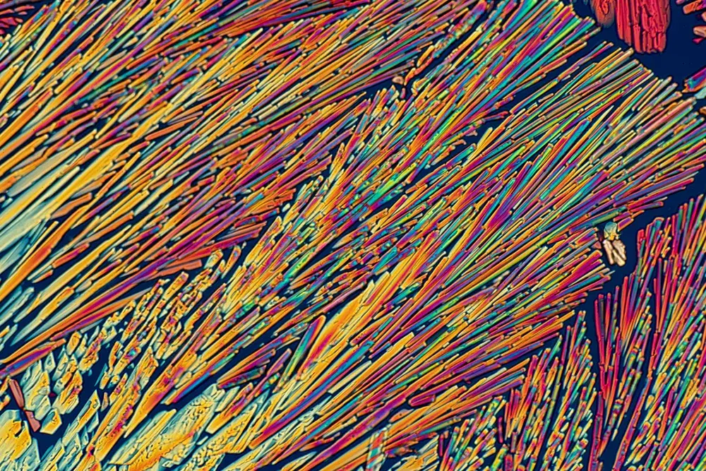
The images begin with crystallography — dissolving a substance, then coaxing it back into crystal form. Salt is the simplest example: dissolve a spoonful in water, let it dry, and you've grown crystals. But dried salt is colourless. The colour comes from somewhere else.
It comes from birefringence — the way certain crystals bend light as it passes through them. The bending splits and shifts the light, and where it does, colour appears. Under a polarized-light microscope, an ordinary compound becomes a field of colour that exists nowhere else.
An analogy
Picture a field of grass standing straight up. Pour water over it and most runs straight to the soil. Now imagine the wind lays the grass flat on its side — pour the water again, and it can no longer fall straight down. It splits, and scatters, and finds a dozen paths.
Light through a birefringent crystal behaves the same way. The crystal's atomic structure bends and splits it — and the result, caught through the microscope, is art.
The process
The difficult substances are the point.
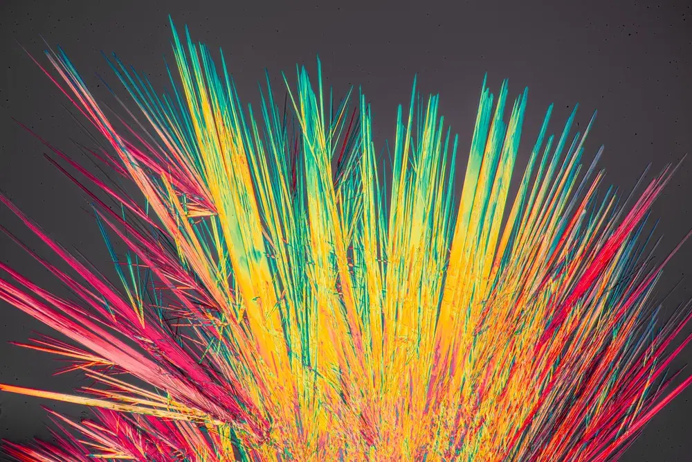
Some materials crystallize willingly. Most do not. Many take extraordinary time, patience, and a scientist's hand to coax into form at all. Salt is easy, and pretty enough — but the work worth making lies past it.
So the collections reach for the difficult and the unexpected: a cocktail, a vitamin, a supplement, a beauty serum. The things you live alongside every day, shown as you have never seen them.
The story
The Micrographer
Micrographer is the work of Matt Femia, who spent seven years in academic and cancer research across Cornell, William & Mary, and Memorial Sloan Kettering Cancer Center in New York — logging over four thousand hours at the microscope, contributing to published work in the field's leading journals and to new immunotherapy treatments for breast-cancer patients.
The first prints were made in a shoebox Manhattan apartment, on a microscope he built himself — 3D-printed parts paired with salvaged optics from medical scopes of the 1980s. A handful of those earliest images circulated through the city. In 2021, Micrographer became a practice of its own.
“These prints are rooted in the memories of my own life, and I hope they find meaning in yours. If nothing else, share in the quiet joy of seeing the world that sits just beneath our eyes.”